Pancreas facts for kids
Quick facts for kids Pancreas |
|
|---|---|
 |
|
| Anatomy of the pancreas | |
| Latin | Pancreas |
| Artery | Inferior pancreaticoduodenal artery, anterior superior pancreaticoduodenal artery, posterior superior pancreaticoduodenal artery, splenic artery |
| Vein | Pancreaticoduodenal veins, pancreatic veins |
| Nerve | Pancreatic plexus, celiac ganglia, vagus nerve |
| Lymph | Splenic lymph nodes, celiac lymph nodes and superior mesenteric lymph nodes |
| Precursor | Pancreatic buds |
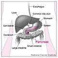
The pancreas is an organ that makes hormones and enzymes to help digestion. The pancreas helps break down carbohydrates, fats, and proteins. The pancreas is behind the stomach and is on the left side of the human body.
The part of the pancreas that makes hormones is called the Islets of Langerhans. The Islets of Langerhans are a small part (2%) of the total cells in the pancreas. The Islets of Langerhans change which chemical they make depending on how much of other chemicals are already in the blood. So, the pancreas works to keep the level of chemicals in balance in the body. If the Islets of Langerhans stop working, a person will suffer from a disease called diabetes. Doctors are experimenting with taking the Islets of Langerhans cells from a donor body and putting them into the pancreas of a person with diabetes to make that person well.
The pancreas belongs to two systems of the body: the digestive system for its role in breaking down nutrients, and the endocrine system for producing hormones.
Hormones
The pancreas releases these hormones:
- Insulin (which decreases the amount of glucose or sugar in the blood)
- Glucagon (which increases the amount of glucose in the blood)
- Somatostatin (which reduces production of insulin and glucagon)
Digestive enzymes
The pancreas releases many different enzymes to help digestion:
- Lipase (which breaks down fats)
- Amylase (which breaks down carbohydrates)
- Trypsinogen and Chymotrypsin (which break down proteins)
- Erepsin, which digests peptones into amino acids.
Structure
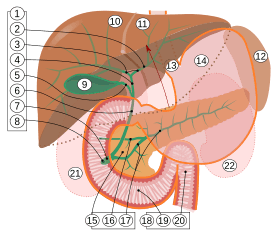
9. Gallbladder, 10–11. Right and left lobes of liver. 12. Spleen.
13. Esophagus. 14. Stomach. 15. Pancreas: 16. Accessory pancreatic duct, 17. Pancreatic duct.
18. Small intestine: 19. Duodenum, 20. Jejunum
21–22. Right and left kidneys.
The front border of the liver has been lifted up (brown arrow).
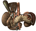
The pancreas is an organ that in humans lies in the upper left part of the abdomen. In adults, it is about 12–15 centimetres (4.7–5.9 in) long, lobulated, and salmon-coloured in appearance.
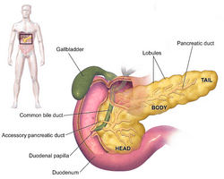
Anatomically, the pancreas is divided into a head, neck, body, and tail. The pancreas stretches from the inner curvature of the duodenum, where the head surrounds two blood vessels, the superior mesenteric artery, and vein. The longest part of the pancreas, the body, stretches across behind the stomach, and the tail of the pancreas ends adjacent to the spleen.
Two ducts, the main pancreatic duct and a smaller accessory pancreatic duct, run through the body of the pancreas, joining with the common bile duct near a small ballooning called the ampulla of Vater. Surrounded by a muscle, the sphincter of Oddi, this opens into the descending part of the duodenum.
Shape
The head of the pancreas sits within the curvature of the duodenum, and wraps around the superior mesenteric artery and vein. To the right sits the descending part of the duodenum, and between these travel the superior and inferior pancreaticoduodenal arteries. Behind rests the inferior vena cava, and in front sits the peritoneal membrane and the transverse colon.
From the back of the head emerges a small uncinate process, which extends to the back of the superior mesenteric vein and ends at the superior mesenteric artery. The superior mesenteric artery passes down in front of the left half across the uncinate process; the superior mesenteric vein runs upward on the right side of the artery and, behind the neck, joins with the lienal vein to form the portal vein.
The body of the pancreas travels from the head, separated by a short neck. The neck is about 2 cm (0.79 in) wide, and sits in front of the portal vein. The gastroduodenal artery and the anterior superior pancreaticoduodenal arteries travel in front of the gland and begin where the neck meets the head of the pancreas. The neck lies mostly behind the pylorus of the stomach.
The body is the largest part of the pancreas, and mostly lies behind the stomach. It has a triangular cross-section, with the tip near the top of the pancreas, and the base at the bottom. The peritoneum sits on top of the front-facing and lower surfaces of the pancreas. Behind the pancreas are several blood vessels, including the aorta, the splenic vein, and the left renal vein, as well as some of the superior mesenteric artery. Below the body of the pancreas sits some of the small intestine, specifically the last part of the duodenum and the jejunum to which it connects, as well as the suspensory ligament of the duodenum which falls between these two. In front of the pancreas sits the transverse colon.
The pancreas narrows towards the tail, which sits next to the spleen. It is usually between 1.3–3.5 cm (0.51–1.38 in) long, and sits between the layers of the ligament between the spleen and the left kidney. The splenic vein, which also passes behind the body of the pancreas, passes behind the tail of the pancreas.
Blood supply
The pancreas has a rich blood supply, with vessels originating as branches of both the coeliac artery and superior mesenteric artery. The splenic artery runs along the top margin of the pancreas, and supplies the left part of the body and the tail of the pancreas through its pancreatic branches, the largest of which is called the greater pancreatic artery. The superior and inferior pancreaticoduodenal arteries run along the anterior and posterior surfaces of the head of the pancreas at its border with the duodenum. These supply the head of the pancreas. These vessels originate join together (anastamose) in the middle.
The body and neck of the pancreas drain into the splenic vein, which sits behind the pancreas. The head drains into, and wraps around, the superior mesenteric and portal veins. The pancreas drains into lymphatic vessels that travel alongside its arteries. These lymphatic vessels drain primarily into pancreaticosplenic nodes, and some into the lymph nodes that lie in front of the aorta.
Images for kids
-
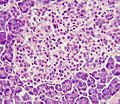
This image shows a pancreatic islet when pancreatic tissue is stained and viewed under a microscope. Parts of the digestive ("exocrine") pancreas can be seen around the islet, more darkly. These contain hazy dark purple granules of inactive digestive enzymes (zymogens).
-
A pancreatic islet that uses fluorescent antibodies to show the location of different cell types in the pancreatic islet. Antibodies against glucagon, secreted by alpha cells, show their peripheral position. Antibodies against insulin, secreted by beta cells, show the more widespread and central position that these cells tend to have.
-
The pancreas originates from the foregut, a precursor tube to part of the digestive tract, as a dorsal and ventral bud. As it develops, the ventral bud rotates to the other side and the two buds fuse together.
-
The pancreas has a role in digestion, highlighted here. Ducts in the pancreas (green) conduct digestive enzymes into the duodenum. This image also shows a pancreatic islet, part of the endocrine pancreas, which contains cells responsible for secretion of insulin and glucagon.
See also
 In Spanish: Páncreas para niños


