Intestinal villus facts for kids
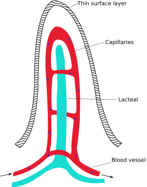
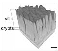
Intestinal villi (singular: villus) are small, finger-like projections that extend into the small intestine. Each villus is approximately 0.5–1.6 mm in length (in humans), and has many microvilli. Each of these microvilli are about 1 µm in length, around 1000 times shorter than a single villus. The intestinal villi are much smaller than any of the circular folds in the intestine.
Villi increase the internal surface area of the intestinal walls making available a greater surface area for absorption. An increased absorptive area is useful because digested nutrients pass into the the villi through diffusion, which is effective only at short distances. In other words, increased surface area decreases the average distance travelled by nutrient molecules, so the effectiveness of diffusion increases. The villi are connected to the blood vessels so the circulating blood then carries these nutrients away.
Images for kids
-
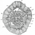
Transverse section of a villus, from the human intestine. X 350. a. Basement membrane, here somewhat shrunken away from the epithelium. b. Lacteal. c. Columnar epithelium. d. Its striated border. e. Goblet cells. f. Leucocytes in epithelium. f’. Leucocytes below epbithelium. g. Blood vessels. h. Muscle cells cut across.
-
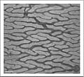
Cross-section histology of small intestinal villi of the human terminal ileum.
-
Microvilli (shaggy hair) show electron dense plaques (open arrow) at their apices.
See also
 In Spanish: Vellosidad intestinal para niños


