Progeria facts for kids
Quick facts for kids Progeria |
|
|---|---|
| Synonyms | Hutchinson–Gilford progeria syndrome (HGPS), progeria syndrome |
 |
|
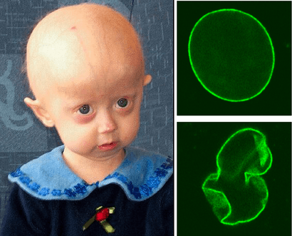
| A young girl with progeria (left). A healthy cell nucleus (right, top) and a progeric cell nucleus (right, bottom). | |
| Symptoms | Growth delay, short height, small face, hair loss |
| Complications | Heart disease, stroke, hip dislocations |
| Usual onset | 9–24 months |
| Causes | Genetic |
| Diagnostic method | Based on symptoms, genetic tests |
| Similar conditions | Hallermann–Streiff syndrome, Gottron's syndrome, Wiedemann–Rautenstrauch syndrome |
| Treatment | Mostly symptomatic |
| Medication | Lonafarnib |
| Prognosis | Average age of death is 13 years |
| Frequency | Rare: 1 in 18 million |
Progeria is an extremely rare autosomal dominant genetic disorder in which symptoms resembling aspects of aging are manifested at a very early age. Progeria is one of several progeroid syndromes. Those born with progeria typically live to their mid-teens to early twenties. It is a genetic condition that occurs as a new mutation, and is rarely inherited, as carriers usually do not live to reproduce children. Although the term progeria applies strictly speaking to all diseases characterized by premature aging symptoms, and is often used as such, it is often applied specifically in reference to Hutchinson–Gilford progeria syndrome (HGPS).
Progeria was first described in 1886 by Jonathan Hutchinson. It was also described independently in 1897 by Hastings Gilford. The condition was later named Hutchinson–Gilford progeria syndrome. The word progeria comes from the Greek words "pro" (πρό), meaning "before" or "premature", and "gēras" (γῆρας), meaning "old age". Scientists are interested in progeria partly because it might reveal clues about the normal process of aging.
Images for kids
-
Ultrastructural analysis of the nuclear envelope in fibroblasts from a subject with HGPS. Low magnification transmission electron microscopic image of a passage 10 PT001 nucleus showed several herniations (a). Two higher-magnification images of the same nucleus at sites of blebs (b and c) showed a close apposition of the chromatin to the nuclear envelope. In a, b, and c, the nucleus is to the left. Scale bars correspond to 2 μm in panel a, and 500 nm in panels b and c.
See also
 In Spanish: Progeria para niños

