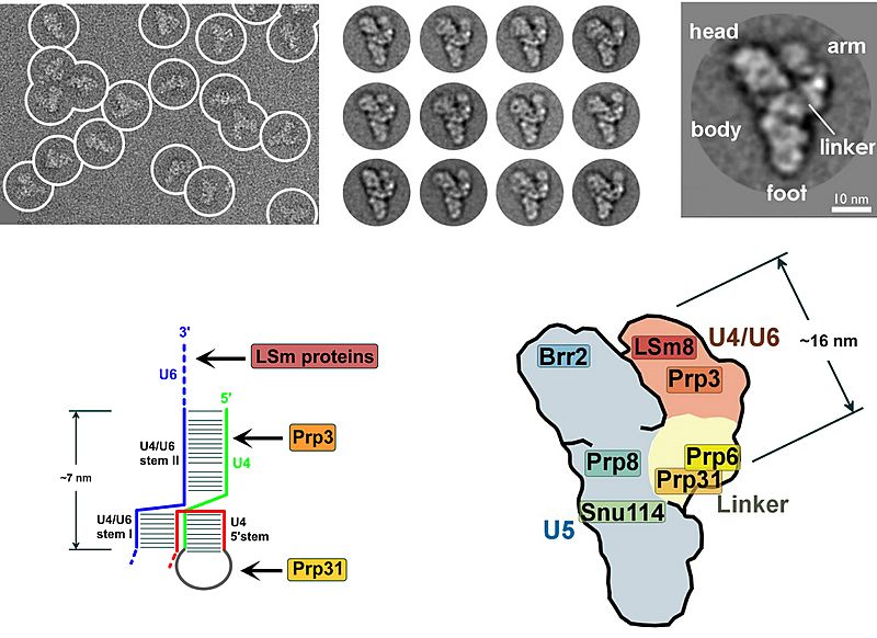
Image: Yeast tri-snRNP

Size of this preview: 800 × 582 pixels. Other resolutions: 320 × 233 pixels | 2,763 × 2,011 pixels.
Original image (2,763 × 2,011 pixels, file size: 1.06 MB, MIME type: image/jpeg)
Description: Above are electron microscopy fields of negatively stained tri-snRNPs. Below left is a schematic of the helices of stem II and the U4 stem-loop of the U4/U6 snRNA duplex. Below right is a cartoon model of the yeast tri-snRNP with shaded areas corresponding to U5 (gray), U4/U6 (orange) and the linker region (yellow).
Title: Yeast tri-snRNP
Credit: Dr Berthold Kastner (Transferred from en.wikipedia to Commons by Vojtech.dostal.)
Author: Berthold Kastner
Usage Terms: GNU Free Documentation License
License: GFDL
License Link: http://www.gnu.org/copyleft/fdl.html
Attribution Required?: Yes
Image usage
The following 2 pages link to this image:

All content from Kiddle encyclopedia articles (including the article images and facts) can be freely used under Attribution-ShareAlike license, unless stated otherwise.
