
Image: Part of whole slide image of a Wilms' tumor of the kidney

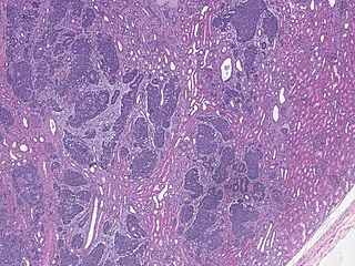
Description: Low magnification micrograph of a Wilms' tumour infiltrating the renal parenchyma. The image is part of a whole slide scan. H&E stain. Wilms tumour is a type of kidney cancer that is seen predominantly in children. The images show the characteristic three components: Malignant small round (blue) cells ~ 2x the size of resting lymphocyte (blastema component). Tubular structures/rosettes (epithelial component). Loose paucicellular stroma with spindle cells (stromal component), seen as grey areas between the above two components In addition, there is surrounding renal tissue, seen as more eosinophilic (pink) areas compared to the stromal tumor component.
Title: Part of whole slide image of a Wilms' tumor of the kidney
Credit: Own work
Author: Mikael Häggström, M.D. Author info - Reusing images - Conflicts of interest: NoneMikael Häggström, M.D. Consent note: Consent from the patient or patient's relatives is regarded as redundant, because of absence of identifiable features (List of HIPAA identifiers) in the media and case information (See also HIPAA case reports guidance).
Usage Terms: Public domain
License: Public domain
Attribution Required?: No
Image usage
The following page links to this image:
