
Image: Histopathology of pulmonary congestion and siderophages
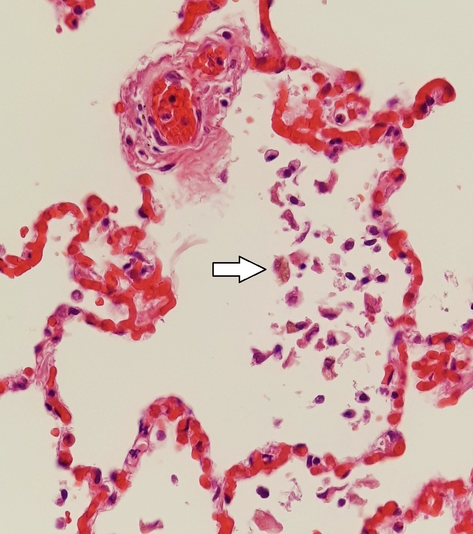
Description: Histopathology showing: - Pulmonary congestion: red blood cells distending pulmonary vessels, and filling alveolar walls - Siderophages: one indicated by white arrow The picture indicates chronic left heart failure. The lymphocytic infiltrate is consistent with this.
Title: Histopathology of pulmonary congestion and siderophages
Credit: Own work
Author: Mikael Häggström, M.D. - Author info - Reusing images Consent from the patient or patient's relatives is regarded as redundant, because of absence of identifiable features (List of HIPAA identifiers) in the media and case information (See also HIPAA case reports guidance).
Usage Terms: Creative Commons Zero, Public Domain Dedication
License: CC0
License Link: http://creativecommons.org/publicdomain/zero/1.0/deed.en
Attribution Required?: No
Image usage
The following page links to this image:
