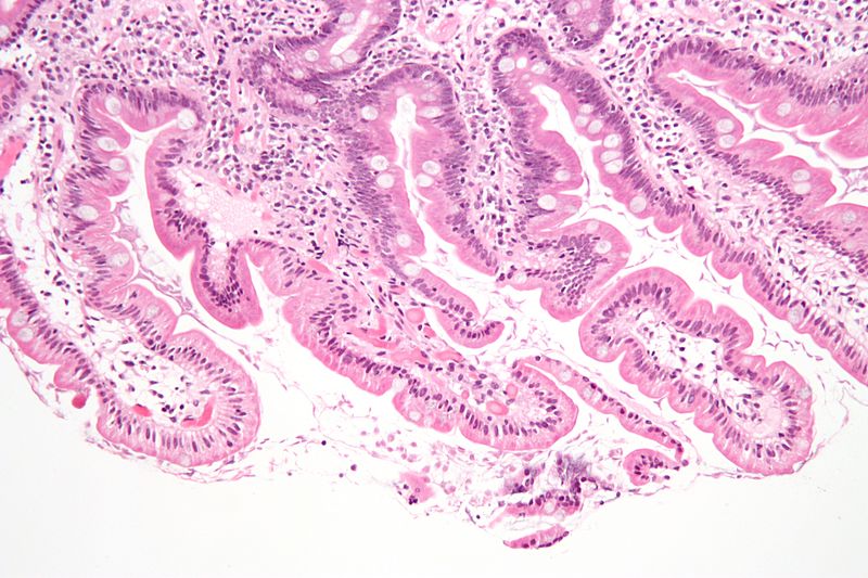
Image: Giardiasis duodenum low

Description: Intermediate magnification micrograph of a small bowel mucosa (duodenum) biopsy with giardiasis. H&E stain. Giardiasis is due to the flagellate protozoan Giardia lamblia. The Giardia organisms are seen in the bowel lumen (bottom of image) and are: Pale staining/translucent. Size: 12-15 micrometers (long axis) x 6-10 micrometers (short axis) -- if seen completely. Most of the Giardia organisms seen are semicircular or crescent shaped -- as the long axis of the organism is rarely in the plane of the (histologic) section. The villi have interepithelial lymphocytes. See also Image:Giardiasis_duodenum_high.jpg - higher magnification view.
Title: Giardiasis duodenum low
Credit: Own work
Author: Nephron
Usage Terms: Creative Commons Attribution-Share Alike 3.0
License: CC BY-SA 3.0
License Link: http://creativecommons.org/licenses/by-sa/3.0
Attribution Required?: Yes
Image usage
The following page links to this image:

