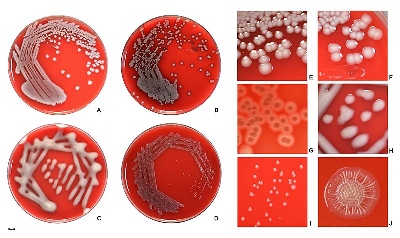
Image: Escherichia coli on agar

Description: Escherichia coli colonies on sheep blood agar after 24 h at 37°C. Fig. A, B: Most strains of E.coli produce smooth, circular, low-convex colonies with entire edge that are about 3-4 mm in diameter. They are greyish, butyrous and readily emulsified. Fig. B, G: Partial digestion of erythrocytes may cause more or less profound discoloration of agar under colonies and in their vicinity (Fig. F). Isolates from urinary tract are quite often beta-hemolytic. Fig. C, H: Less common are higly mucoid strains, typically isolated from urine. Fig. D, I: or small-colony variants, previously known as dwarf colonies, typically isolated from urine. Fig. J: urinary isolate - Rough colonies are quite uncommon in clinical samples
Title: Escherichia coli on agar
Credit: Own work
Author: HansN.
Usage Terms: Creative Commons Attribution-Share Alike 4.0
License: CC BY-SA 4.0
License Link: https://creativecommons.org/licenses/by-sa/4.0
Attribution Required?: Yes
Image usage
The following page links to this image:

