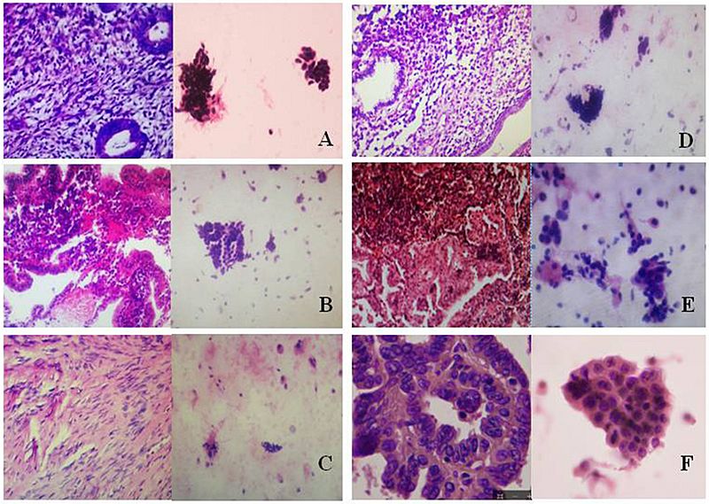
Image: Endometrial histopathologies and cytopathologies

Description: Histopathologic and cytopathologic images. (A) proliferative endometrium (Left: HE × 400) and proliferative endometrial cells (Right: HE × 100) (B) secretory endometrium (Left: HE × 10) and secretory endometrial cells (Right: HE × 10) (C) atrophic endometrium (Left: HE × 10) and atrophic endometrial cells (Right: HE × 10) (D) mixed endometrium (Left: HE × 10) and mixed endometrial cells (Right: HE × 10) (E): endometrial atypical hyperplasia (Left: HE × 10) and endometrial atypical cells (Right: HE × 200) (F) endometrial carcinoma (Left: HE × 400) and endometrial cancer cells (Right: HE × 400).
Title: Endometrial histopathologies and cytopathologies
Credit: (2019). "An Efficacious Endometrial Sampler for Screening Endometrial Cancer". Frontiers in Oncology 9. DOI:10.3389/fonc.2019.00067. ISSN 2234-943X. - "This is an open-access article distributed under the terms of the Creative Commons Attribution License (CC BY)."
Author: Lu Han, Jiang Du, Lanbo Zhao, Chao Sun, Qi Wang, Xiaoqian Tuo, Huilian Hou, Yu Liu, Qing Wang, Qurat Ulain, Shulan Lv2, Guanjun Zhang, Qing Song and Qiling Li
Usage Terms: Creative Commons Attribution 4.0
License: CC BY 4.0
License Link: https://creativecommons.org/licenses/by/4.0
Attribution Required?: Yes
Image usage
The following page links to this image:

