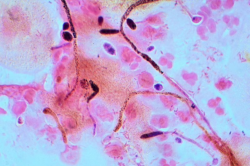
Image: Candida Gram stain

Description: Candida albicans from a vaginal swab. This is a high resolution (within the constraints of optical microscopy) micrograph of an important pathogen. The magnification of the original NEF image is around 1,000 times. On most PC screens it will be around 5,000 times, but much of this will be dead magnification. The image shows the hyphae (filaments) and the oval and elliptical chlamydospores stained blue-black. The pink blobs are vaginal epithelial cells and the dark granules are common artefacts of the Gram-stain. (Note: microscope lenses do not have F-numbers they have Numerical apertures, this one was NA 1.25 and oil immersion).
Title: Candida Gram stain
Credit: Own work
Author: Graham Beards
Usage Terms: Creative Commons Attribution-Share Alike 4.0
License: CC BY-SA 4.0
License Link: https://creativecommons.org/licenses/by-sa/4.0
Attribution Required?: Yes
Image usage
The following page links to this image:

