Image: Astrocyte5

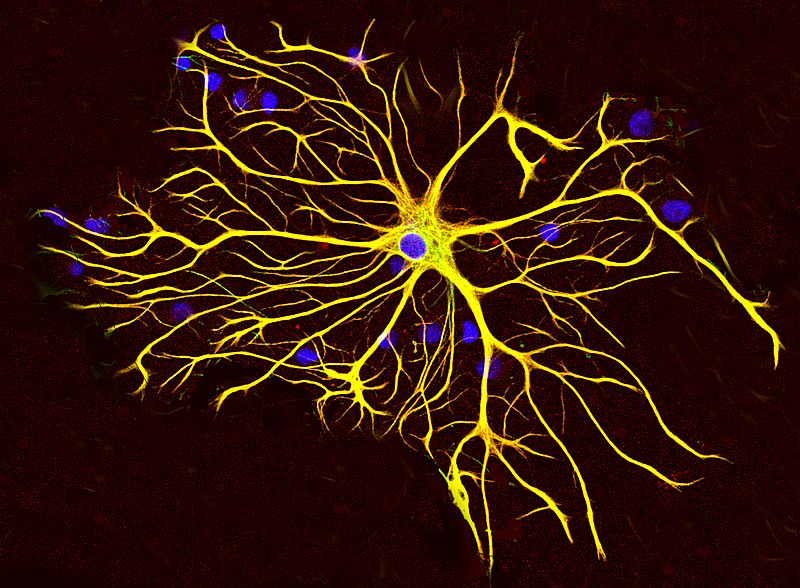
Description: An astrocyte cell grown in tissue culture stained with antibodies to GFAP and vimentin. The GFAP is coupled to a red fluorescent dye and the vimentin is coupled to a green fluorescent dye. Both proteins are present in large amounts in the intermediate filaments of this cell, so the cell appears yellow, the result of combining strong red and green signals. The blue signal is DNA revealed with DAPI, and shows the nucleus of the astrocyte and of other cells in this image. Image was captured on a confocal microscope in the EnCor Biotechnology laboratory.
Title: Astrocyte5
Credit: Own work
Author: GerryShaw
Usage Terms: Creative Commons Attribution-Share Alike 3.0
License: CC BY-SA 3.0
License Link: http://creativecommons.org/licenses/by-sa/3.0
Attribution Required?: Yes
Image usage
The following page links to this image:

