
Image: Assembly of fibrous muscle, fat, and vascular tissues to cultured steak.webp
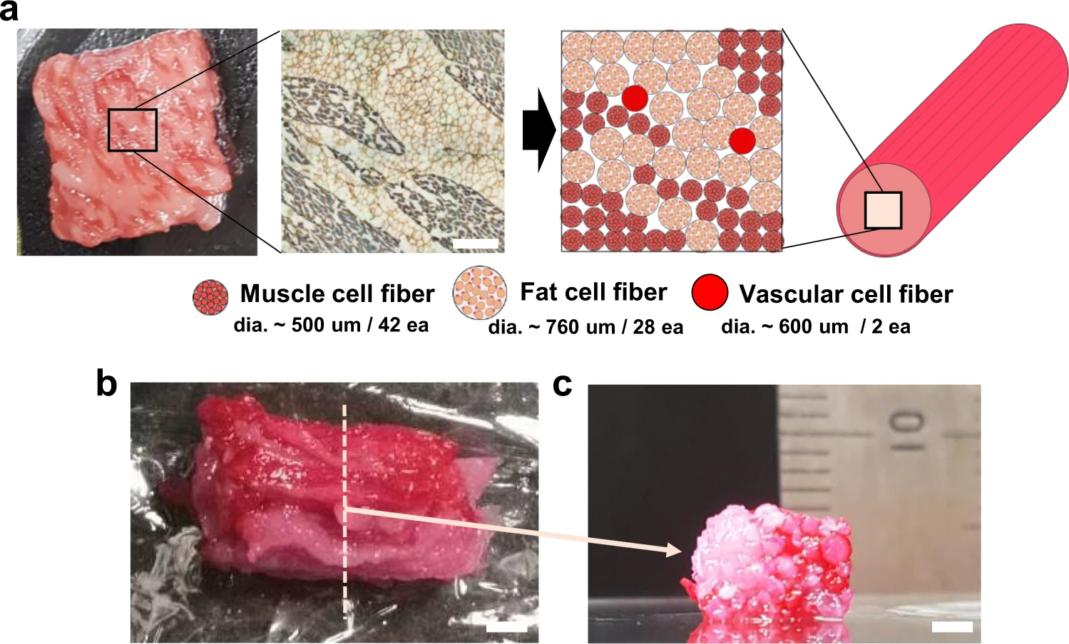
Description: "a Assembly schematic- (right) based sarcomeric α-actinin (blue) and laminin- (brown) stained image (left) of the commercial meat. It is assumed that the diameters of the fibrous muscle, fat, and vascular tissues are about 500, 760, and 600 µm, respectively. Scale bar, 1 mm. b, c Optical images of the cultured steak by assembling muscle (42 ea.), fat (28 ea.), and vascular (2 ea.) tissues at (b) the top and (c) cross-section view of the dotted-line area. Muscle and vascular tissue were stained with carmine (red color), but fat tissue was not. Scale bars, 2 mm."
Author: Authors of the study: Dong-Hee Kang, Fiona Louis, Hao Liu, Hiroshi Shimoda, Yasutaka Nishiyama, Hajime Nozawa, Makoto Kakitani, Daisuke Takagi, Daijiro Kasa, Eiji Nagamori, Shinji Irie, Shiro Kitano & Michiya Matsusaki
Usage Terms: Creative Commons Attribution-Share Alike 3.0
License: CC-BY-SA-3.0
License Link: http://creativecommons.org/licenses/by-sa/3.0/
Attribution Required?: Yes
Image usage
The following page links to this image:
