Image: Anchiornis plumage

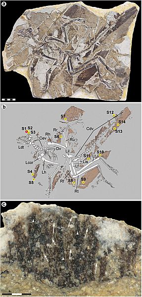
Description: Anchiornis huxleyi specimen YFGP-T5199. (a) Photographic and (b) diagrammatic representation. Numbered circles denote location of plumage samples used for molecular and/or imaging analyses. Red circle (S1) demarcates the ‘forecrown’ sample used as the basis for our investigation; yellow circles (S2–S14) indicate samples used for supportive SEM imaging. Cdv, caudal vertebrae; Cev, cervical vertebrae; Dv, dorsal vertebrae; Lcor, left coracoid; Ldt, left dentary; Lf, left femur; Lh, left humerus; Lil, left ilium; Lis, left ischium; Lt, left tibia; Rf, right femur; Rh, right humerus; Rr, right radius; Rt, right tibia; Ru, right ulna. Scale bar: 5 cm. Photograph by Pascal Godefroit and Ulysse Lefèvre. Drawing by Ulysse Lefèvre. (c) Detail of S1 after initial preparation showing darker central strands (arrowheads) with diffuse arrays of filaments branching laterally at acute angles (arrows). Note that the analysed area is still covered by sedimentary matrix (see also Supplementary Fig. S1). Scale bar: 300 μm. Photograph by Johan Lindgren.
Title: Anchiornis plumage
Credit: http://www.nature.com/articles/srep13520
Author: Johan Lindgren, Peter Sjövall, Ryan M. Carney, Aude Cincotta, Per Uvdal, Steven W. Hutcheson, Ola Gustafsson, Ulysse Lefèvre, François Escuillié, Jimmy Heimdal, Anders Engdahl, Johan A. Gren, Benjamin P. Kear, Kazumasa Wakamatsu, Johan Yans & Pascal Godefroit
Usage Terms: Creative Commons Attribution 4.0
License: CC BY 4.0
License Link: http://creativecommons.org/licenses/by/4.0
Attribution Required?: Yes
Image usage
The following page links to this image:

