Image: 42003 2019 308 Fig2 HTML

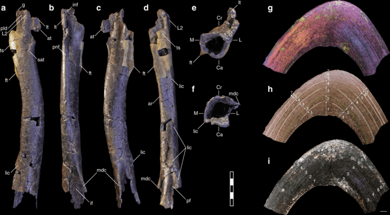
Description: Right femur of M. intrepidus (NCSM 33392). (a) Lateral, (b) cranial, (c) medial, (d) caudal, (e) proximal, and (f) distal views. Partial mid-diaphyseal cross-section of the femur shown in (g) polarized light with lambda filter, (h) natural light with numbered arrows and tracings indicating seven growth cycles (see Supplementary Fig. 5), and (i) polarized light. Abbreviations: ar adductor ridge, at accessory trochanter, Ca caudal aspect, Cr cranial aspect, ft fourth trochanter, if intercondylar fossa, inf intertrochanteric nutrient foramen, L lateral aspect, L2 lobe on lesser trochanter (sensu17), lic linea intermuscularis caudalis. lt lesser trochanter, M medial aspect, mdc mesiodistal crest, pf popliteal fossa, pld lateral depression, proximal. pnf principle nutrient foramen, sat semicircular accessory tuberosity, ts trochanteric shelf. Scale bar (a–e) 5 cm; (g–i) 1 mm
Title: 42003 2019 308 Fig2 HTML
Credit: https://www.nature.com/articles/s42003-019-0308-7#MOESM1
Author: Lindsay E. Zanno, Ryan T. Tucker, Aurore Canoville, Haviv M. Avrahami, Terry A. Gates & Peter J. Makovicky
Usage Terms: Creative Commons Attribution 4.0
License: CC BY 4.0
License Link: https://creativecommons.org/licenses/by/4.0
Attribution Required?: Yes
Image usage
The following page links to this image:

