Image: 3phv HIV-prot rib

Size of this preview: 755 × 600 pixels. Other resolutions: 302 × 240 pixels | 2,400 × 1,907 pixels.
Original image (2,400 × 1,907 pixels, file size: 200 KB, MIME type: image/png)
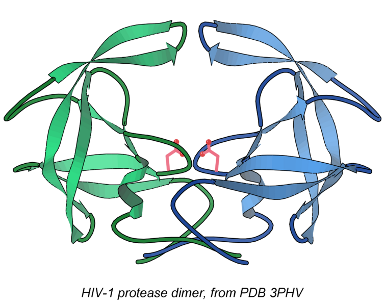
Description: Ribbon schematic of the HIV-1 protease dimer, with the two chains in green and blue and the active-site Asp sidechains in pink. The "flaps" that cover the substrate or drug binding site are at the top, and the intertwined beta structure that forms the dimer is at bottom. Image made in KiNG, from PDB file 3PHV.
Title: 3phv HIV-prot rib
Credit: Own work
Author: Dcrjsr
Usage Terms: Creative Commons Attribution 4.0
License: CC BY 4.0
License Link: https://creativecommons.org/licenses/by/4.0
Attribution Required?: Yes
Image usage
The following page links to this image:

All content from Kiddle encyclopedia articles (including the article images and facts) can be freely used under Attribution-ShareAlike license, unless stated otherwise.
